
Imaging in the second near-infrared window (NIR-II, 1000-1700 nm) is promising in biomedical basic research and clinical translation due to advantages of low tissue absorption and scattering properties, deep tissue penetration, and improved spatial resolution.
However, real-time NIR-II fluorescence imaging of a living body with high resolution still faces enormous challenges because of the inherent shortcomings of fluorescence imaging including small Stokes shift and short optical imaging lifetime.
Recently, a collaborative research team has developed a NIR-II phosphorescent imaging strategy based on the novel phosphorescent probe.
The team includes Prof. HU Zhenhua from the Institute of Automation of the Chinese Academy of Sciences, Profs. CHANG Baisong and SUN Taolei from the Wuhan University of Technology, Prof. CHENG Zhen from Stanford University and Shanghai Institute of Materia Medica of the Chinese Academy of Sciences.
The study was published in Nature Biomedical Engineering on August 12.
The researchers developed specific CISe nanotubes for NIR-II phosphorescent imaging with an emission peak of 1130 nm. The excited-state relaxation procedure of the CISe nanotubes was modified through H-bonding-driven reversible self-limited assembly, which prolonged the lifetime of the emitted light from 73 ns to 336 μs.
Based on the fluorescence-phosphorescence transition mechanism of CISe nanotubes, a novel phosphorescent imaging method was then developed. Specific imaging capabilities for tumors were demonstrated by in vivo real-time imaging of living mice, with the background interference from normal tissues significantly suppressed.
"After injection of the nanoprobe into the tail vein for 24 h, the signal ratio of tumor-to-normal tissue reached as high as 190, and the resolution of blood vessels increased up to 7.6 μm, showing extraordinary imaging performance in living animals," said Prof. HU. This method has exhibited extensive prospects in biomedical research.
In their previous study, Prof. TIAN Jie, Prof. HU Zhenhua and their collaborators conducted the first-in-human clinical translation of NIR-II fluorescence imaging, and more primary and metastatic liver tumors were detected compared with traditional imaging techniques.
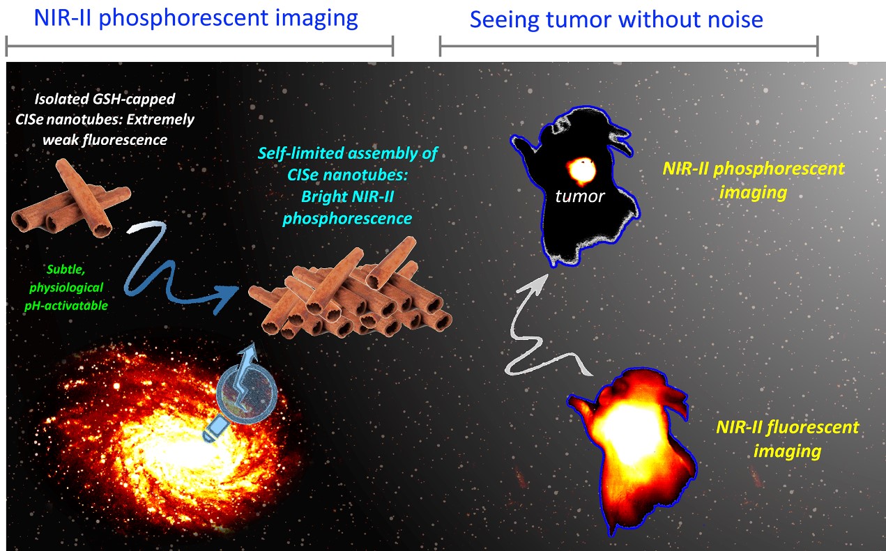
High performance and specific imaging of tumors using CISe NIR-II phosphorescent probe (Image by CASIA)
Contact:
ZHANG Xiaohan, PIO, Institute of Automation, Chinese Academy of Sciences
Email: xiaohan.zhang@ia.ac.cn